/* Template Name: PageOfPosts */ Angie's CPD » Reflectives

Colonic Stenting 29/03/11 Year 4

March 29th, 2011

Description

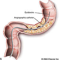
Schematic drawing of a tumorous lesion in the rectosigmoid junction traversed with a guidewire and catheter. C, Terumo guidewire (left) and angiographic catheters (right), Cobra and Headhunter types. D, Schematic drawing of the stent instrument advanced over the superstiff wire and the released stent in place.

Imaging consult colonic stenting.jpg


Week 13 Year 4

March 21st, 2011

21 March 2011

This was my final week of placement as a student. It was quite daunting as I was very conscious of the fact that the next time I work in a radiography department I will be a qualified radiographer. It was also very exciting as, after 5 years, I can now see the end of the course in sight.
During the week I carried out a general practitioner (GP) request on a female patient who had presented to her GP with pain and tenderness in her forearm. The clinical history was brief so I asked the patient for more details. She showed the area of the pain which looked a little swollen but nothing remarkable. She informed me there had been no trauma and she didn’t understand why it was painful. I then continued with the examination of an AP and lateral forearm.
On processing the image it was very clear there was something obviously amiss with the patients’ ulnar. It looked as if something had taken a clean bite out of the patients’ bone, I had seen a similar case previously and thought it looked like osteosarcoma. I asked another radiographer for her opinion and we decided to get a radiologist to have a look before letting the patient leave. It was the opinion of the radiologist that the patient did have osteosarcoma and he was going to telephone her GP straight away so she could get an urgent referral to the osteosarcoma team.
I found the patients symptoms for this very surprising and I could understand why it took her so long to visit her doctor. The patient was 32 years of age with an 18 month old baby so it was understandable that she first thought she may have bumped it without realising and it would get better.
Osteosarcoma is a primary malignant tumour of bone and the cause of it is unknown. According to Shenoy at el (2007) osteosarcoma is the most common malignant bone tumour which conflicts with the opinion of Pretorius and Jeffrey (2006) who state osteosarcoma as being the second most common primary bone tumour after multiple myeloma. It is also the opinion of Pretorius and Jeffrey (2006) that sarcomas tend to undergo hematogenous spread, with pulmonary metastases being the most common.
According to Wang and Houston (2005) most osteosarcomas are sporadic, whereas inherited predisposition accounts for a small number of cases, along with this 20% of patients with osteosarcoma present clinically with overt metastatic disease, with the presence of metastatic disease being a strong predictor of a poor outcome. The disease is believed to originate from primitive mesenchymal cells but also may arise from pluripotential mesenchymal cells. Osteosarcoma is a deadly cancer and is commonly fatal when it metastasizes to the lungs. It usually forms as a single lesion on long bones, with the distal femur being the most common, then the proximal humerus and proximal tibia.
Several tests are necessary to diagnose osteosarcoma. Initially, a blood test followed by imaging tests to detect any other tumours and their location within the body. This can be performed by an X-ray, MRI, CT scan or bone scan. Confirmation of osteosarcoma can also be done by performing a biopsy to remove a piece of bone tissue.
Treatment for osteosarcoma depends on the location of the tumour and the severity of the illness. Treatments may include neoadjuvant therapy which is the administration of a therapeutic agent before the main treatment of chemotherapy to destroy cancerous cells and prevent the growth of new malignant cells. The aim of neoadjuvant therapy is to reduce the size or extent of the cancer before using radical treatment intervention, thus making procedures easier, more likely to succeed, and reducing the consequences of a more extensive treatment technique that would be required if the tumour wasn’t reduced in size or extent. Additionally, radiation therapy, which uses high-energy radiation beams to shrink tumours and destroy cancer cells may also be used to treat osteosarcoma. In advanced stages of osteosarcoma, doctors may suggest amputation to remove the affected limb. Advances in surgical techniques have allowed a reduction in the number of amputations associated with osteosarcoma. Tumours can now be removed from the affected bone without removing a limb, this is done by using artificial bones or bones from other parts of the body. These advances in surgical techniques to treat osteosarcoma have resulted in higher survival rates, and high limb salvage rates associated with this type of cancer. Attached to this writing are images taken from x-ray 2000 and the childrens specialist website.

Pretorius, E. S. and Jeffrey A. S. 2006. Radiology secrets. 2nd ed. Philadelphia: Mosby Elsevier

Shenoy, R. Pillai, A. Sokhi, K. Porter, D. Ried, R. 2008. Survival trends in osteosarcoma of humerus. European Journal of Cancer Care 17, pp. 261–269.

Wang, L. 2005. Biology of Osteogenic Sarcoma. Cancer Journal. pp. 294-305.

Ehow Health. 2011. How long are treatments for osteosarcoma. [online] Available at: http://www.ehow.com/how-does_5251031_long-treatments-osteosarcoma_.html [Accessed March 27]

Childrens Specialists. 2011. Musculoskeletal Tumors and Infections. [online] Available at: http://www.cssd.us/body.cfm?id=1238 [Accessed March 27]

Week 12 Year 4

March 21st, 2011

This week I was working in the interventional room at the Western General Hospital. There were a number of procedures that were new to me, so it was a very interesting week. Some of the procedures that I assisted with were barium enemas, water soluble enemas, Hickman lines, colonic stenting and gastrostomy tubes. There were a number of Hickman line procedures where I was allowed to perform the screening and also carry out nursing duties. All the patients I assisted with were all receiving their Hickman lines for treatment of different types of cancers.
Hickman lines consist of a soft plastic tube that is tunneled beneath the skin and placed in a large vein. The insertion is carried out for a number of reasons and depending on the requirement of the line will depend on the number of connectors required, which can be single, double or triple lumen catheter. These catheters can then be used to give fluids, drugs and for taking blood samples. This procedure is usually carried out to save the patient repeatedly having to endure needles for giving or providing blood samples or for administering drugs.
The radiologist marked the vein using ultrasound then proceeded to administer a local anaesthetic. He then made two cuts in the patients’ chest, one to tunnel the catheter and the other near the collarbone. The line was then inserted into position with the use of fluoroscopy screening, this allows real time visualisation of the line and the anatomical structure ensuring the line is positioned correctly. Once the Radiologist was happy with the position he then flushed the line to ensure it was working correctly. He then inserted two stitches at either end of the tube insertions to secure the catheter in position and to stop it from moving. Patients will have their Hickman lines in situ for the period of their treatment before having them removed. The use of fluoroscopy substantially decreases the amount of radiation needed to produce clinically useful images (Pretorius and Solomon 2006).

Another procedure that I assisted with was a colonic stenting on an elderly female patient with an abdominal obstruction. The patient had previously been diagnosed with colon cancer and had a 10 centimetre stricture causing an obstruction. The patient had previously been offered surgery to remove the tumour but had refused it as she didn’t want to undergo a major operation.
The procedure was carried out by a radiologist and a gastroenterologist consultant. The gastroenterologist consultant proceeded to guide the colonoscopy into the patients’ colon to try and pass the stricture. The radiologist then proceeded to insert a guide wire beyond the blockage, using fluoroscopy guidance. A small catheter was then positioned over the wire and the guide wire removed. Another wire was then put down the catheter with a balloon and stent on the end which was ready to be expanded when it was in the correct position. Contrast dye was given to show the bowel outline and the exact position of the blockage for positioning of the stent. Once in the correct position the wire was removed leaving the stent in position. According to Dionig at el, (2007) management of colorectal obstruction by using a metallic stent is a safe and effective procedure with good technical and clinical success; the use of stent can prevent the need for surgery in patients with disseminated disease; it can prevent both temporary and permanent stomas and may mitigate the need for emergency operations for colonic obstruction.
This procedure proved to be a difficult procedure as it was difficult for the gastroenterologist to pass the strictures part of the bowel with the procedure taking longer than hoped.
Possible complications with this procedure can be the movement of the stent where it may migrate further down the bowel. This will then cause the stricture to return and result in the patient having to endure removal of the stent with a further second stent having to be positioned. Another complication can be the risk of perforation to the bowel wall.

Pretorius, E. S. and Jeffrey A. S. 2006. Radiology secrets. 2nd ed. Philadelphia: Mosby Elsevier

Dionigi, G. Villa, F. Rovera, F. Boni, L. Carrafiello, G. Annoni, M. Castano, P. Bianchi, V. Mangini, M. Recaldini, C. Lagana, D. Bacuzzi, A. Dionigi, R. 2007. Colonic stenting for malignant disease: Review of literature. Surgical Oncology, 16 pp. 153-155.

Week 11 Year 4

March 14th, 2011

13 March 2011

This week I was in the Royal Hospital for Sick Kids. It was a very interesting week with me experiencing and learning a lot. Paediatric radiography is very different to general adult radiography. Rules and protocols are strictly defined for paediatric radiography and students who are on placement are made aware of them very quickly.
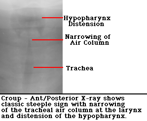
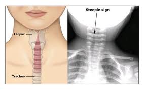
While there I was able to attend the weekly A & E meeting. This meeting allows the A & E consultants and the radiologists to discuss any interesting cases or cases where there are questions surrounding patients’ diagnosis or follow up. Being able to sit in on these meetings, from a student’s point of view, was extremely interesting and very useful. One patient on the list of discussion was a two year old who had ingested a liquid tab designed for washing machines. What made this particularly interesting was a feature on the x-ray that was pointed out and referred to as a steeple sign. They explained this sign is usual in children with croup or children who have experienced near drowning and referred to it as being laryngotracheobronchitis. This steeple sign is a tapering of the trachea which can be seen superiorly on a frontal chest radiograph and is reminiscient of a church steeple.
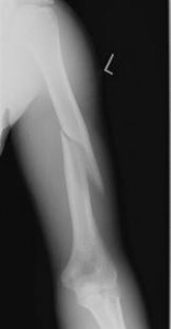
Another case that was discussed was regarding a possible non accidental injury (NAI) of a 7 month old child who had been brought in by his parents due to continued crying and seemed to show signs of a sore arm. It was discovered on x-ray the patient had a spiral fracture of the humerus with minimal displacement. However the fracture travelled from the distal humerus and ran over half way up the shaft of the humerus. It was highlighted by the consultant at the meeting as the patient had been seen previously with a case of severe scabies and also the patient had multiple bruises along with a number of other suspicions which were not discussed. The consultant also questioned the mechanism of injury and said the story did not seem to relate to the extent of the injury. He went on to inform the radiologist that he had reported the case to the child protection team for further investigation.
Later on in the week I was in ITU performing a chest x-ray on an 18 month old patient. The little girl suffered from a rare condition called Jeune syndrome and had previously undergone surgery at Great Ormond Street Hospital to reduce the restriction of her breathing due to her ribs not growing and crushing her lungs. The consultant had informed us that he was extremely concerned for the child and he was unsure if she was going to recover. She had now had a tracheostomy performed to try and assist with her breathing however this didn’t seem to be helping.
An article from emedicine refers to Jeune syndrome as a disorder of bone growth caused by changes in the IFT80 gene. Common signs include a small chest and short ribs which restrict the growth and expansion of the lungs often causing life threatening complications. Other symptoms can include shortened bones in the arms and legs, unusually shaped pelvic bones, and extra fingers and/or toes (polydactyl). Children that survive the difficulties of the breathing and lung challenges at infancy, can then go on to later develop life-threatening kidney problems or heart defects and a narrowing of the airway. Less common features of Jeune syndrome can include liver disease, pancreatic cysts, dental abnormalities, and an eye disease called retinal dystrophy that can lead to vision loss.

Attached to this writing are images on the various topics discussed above.

Children’s Specialists. 2011. Musculoskeletal Tumors and Infections. [online] Available at: http://www.cssd.us/body.cfm?id=1238 [Accessed March 27]
Aswcpoets. 2011. Steeple sign. [online] Available at:http://www.aswcpoets.com/wp-content/uploads/2011/01/croup_steeple_sign.jpg [Accessed March 12]

Radiopaedia. 2011. Croup – steeple sign. [online] Available at:http://radiopaedia.org/cases/croup-steeple-sign [Accessed March 12]

Radiopaedia. 2011. Skeletal dysplasia. [online] Available at:http://radiopaedia.org/articles/skeletal-dysplasia [Accessed March 12]

Dukes Orthopaedics. 2011. Pediatric Supracondylar Fractures of the Humerus [online] Available at: http://www.wheelessonline.com/ortho/pediatric_supracondylar_fractures_of_the_humerus [Accessed March 12]

Emedicine. 2011. Genetics of Asphyxiating Thoracic Dystrophy (Jeune Syndrome)
[online] Available at:http://emedicine.medscape.com/article/945537-overview [Accessed March 12]


?>