Week 5 Endovascular Repair Year 3
This was week 5 of placement and I was at Queen Margaret Hospital. It was my second day of placement and I was in a newly commissioned digital room. I was excited to be using the new equipment; however I was also very intimidated. Not only was there a new computer to learn to understand, I also found that I had to think differently about each examination. I had only just become comfortable with performing most of the examinations in rooms with a Computed Radiography (CR) system using cassettes, so the difference in environment threw me initially. I found in the beginning I was over thinking things especially when it came to performing extremity work. I was so used to using a cassette and thinking about how the image will look on the cassette and moving it to suit the patients’ position. I found myself thinking about how I could position the patient on the digital system so I could still have a good image.
It settled my nerves to know that even the radiographers I was working with were also a bit nervous using the new equipment. Later that afternoon there was a request of an abdomen examination. I had completed a few abdomen requests that day so didn’t have any problem in performing the examination until, that was, I had read the request in full. The request was for an AP, Lateral and Left and Right Anterior Oblique. I informed the radiographer that I wasn’t confident in performing the Lateral and the Left and Right anterior oblique as I had only read about there positioning techniques but had never saw them being performed.
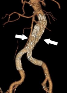
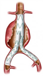
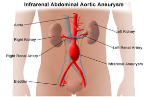
The patient was in for a review due to having an endovascular aneurysm repair. I had never heard of this before and had to ask the radiographer what this was. She explained the patient had a stent placed in his aorta due to an aneurysm which was permanently dilated which is usually caused by a weakening in the vessel wall.
I performed the AP abdomen and the radiographer then performed the others while I observed. The images obtained were great, showing all sides of the stent for the patients’ consultant to review. While observing the images I could see the stent looked like wire mesh which extended from the lower part of the aorta to the upper parts of the common iliac artery, which look like a pair of metal trousers. I found this really interesting and have since done some research into understanding what is involved in endovascular aneurysm repair. I have attached some images and a website address, which shows a good animation of an aneurysm repair, to this piece of writing. This information helped increase my understanding of endovascular aneurysm repair.
This website gives you lots of information and a good understanding of endovascular aneurysm repair, with animations of the procedure.
 Print This Post


