Week 12 Year 4
This week I was working in the interventional room at the Western General Hospital. There were a number of procedures that were new to me, so it was a very interesting week. Some of the procedures that I assisted with were barium enemas, water soluble enemas, Hickman lines, colonic stenting and gastrostomy tubes. There were a number of Hickman line procedures where I was allowed to perform the screening and also carry out nursing duties. All the patients I assisted with were all receiving their Hickman lines for treatment of different types of cancers.
Hickman lines consist of a soft plastic tube that is tunneled beneath the skin and placed in a large vein. The insertion is carried out for a number of reasons and depending on the requirement of the line will depend on the number of connectors required, which can be single, double or triple lumen catheter. These catheters can then be used to give fluids, drugs and for taking blood samples. This procedure is usually carried out to save the patient repeatedly having to endure needles for giving or providing blood samples or for administering drugs.
The radiologist marked the vein using ultrasound then proceeded to administer a local anaesthetic. He then made two cuts in the patients’ chest, one to tunnel the catheter and the other near the collarbone. The line was then inserted into position with the use of fluoroscopy screening, this allows real time visualisation of the line and the anatomical structure ensuring the line is positioned correctly. Once the Radiologist was happy with the position he then flushed the line to ensure it was working correctly. He then inserted two stitches at either end of the tube insertions to secure the catheter in position and to stop it from moving. Patients will have their Hickman lines in situ for the period of their treatment before having them removed. The use of fluoroscopy substantially decreases the amount of radiation needed to produce clinically useful images (Pretorius and Solomon 2006).
Another procedure that I assisted with was a colonic stenting on an elderly female patient with an abdominal obstruction. The patient had previously been diagnosed with colon cancer and had a 10 centimetre stricture causing an obstruction. The patient had previously been offered surgery to remove the tumour but had refused it as she didn’t want to undergo a major operation.
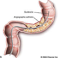
The procedure was carried out by a radiologist and a gastroenterologist consultant. The gastroenterologist consultant proceeded to guide the colonoscopy into the patients’ colon to try and pass the stricture. The radiologist then proceeded to insert a guide wire beyond the blockage, using fluoroscopy guidance. A small catheter was then positioned over the wire and the guide wire removed. Another wire was then put down the catheter with a balloon and stent on the end which was ready to be expanded when it was in the correct position. Contrast dye was given to show the bowel outline and the exact position of the blockage for positioning of the stent. Once in the correct position the wire was removed leaving the stent in position. According to Dionig at el, (2007) management of colorectal obstruction by using a metallic stent is a safe and effective procedure with good technical and clinical success; the use of stent can prevent the need for surgery in patients with disseminated disease; it can prevent both temporary and permanent stomas and may mitigate the need for emergency operations for colonic obstruction.
This procedure proved to be a difficult procedure as it was difficult for the gastroenterologist to pass the strictures part of the bowel with the procedure taking longer than hoped.
Possible complications with this procedure can be the movement of the stent where it may migrate further down the bowel. This will then cause the stricture to return and result in the patient having to endure removal of the stent with a further second stent having to be positioned. Another complication can be the risk of perforation to the bowel wall.
Pretorius, E. S. and Jeffrey A. S. 2006. Radiology secrets. 2nd ed. Philadelphia: Mosby Elsevier
Dionigi, G. Villa, F. Rovera, F. Boni, L. Carrafiello, G. Annoni, M. Castano, P. Bianchi, V. Mangini, M. Recaldini, C. Lagana, D. Bacuzzi, A. Dionigi, R. 2007. Colonic stenting for malignant disease: Review of literature. Surgical Oncology, 16 pp. 153-155.
 Print This Post
