Archive for January, 2011

Week 2 Year 4

Tuesday, January 4th, 2011

This was my second week back at Crosshouse and I had been placed to attend wards and theatres for the week. One particular morning there were two theatre cases which I found to be interesting. The first was a hip arthrogram on a 10 month old girl. The second was for an osteotomy of the hip/femur on an 11 month old female patient. Both had been born with varying degrees of Development Dysplasia of the Hip (DDH).

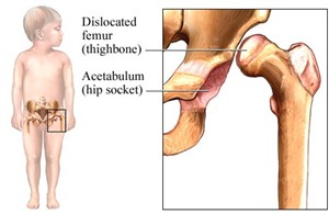
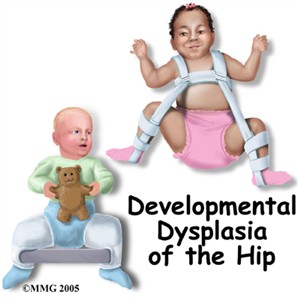
DDH is discovered as part of a routine hip examination check performed on all new born babies. High risk babies are female Caucasian as determined by Pretorius and Solomon (2006). This condition occurs when the formation of the hip joint is dysplastic (abnormal development or growth) or malformed later after birth. The femoral head of the femur and the socket of the acetabulum are made up mostly of cartilage and must be properly orientated for the correct formation. An examination described by Pretorius and Solomon (2006) describes a routine examination on new born babies as the Barlow manoeuvre which dislocates the femoral head rearwards and the Ortolani manoeuvre which reduces the recently dislocated hip usually with a resultant clunk. This examination is carried out for any subluxtion (partially out of alignment) or any instability in the hip joints. Treatments for hip dysplasia depend on the age of the patient. Treatment used for children less than six months of age is a Pavlik harness. The harness holds the hip in an abducted and flexed position. This position allows the best orientation between the femoral head and the acetabulum and allows the hip joint to remodel and develop normally. The harness is then worn full time for six to eight weeks until the hip has stabilised.

The first procedure was an arthrogram of the hip; this was being performed to allow visualisation of the hip joint space and shape. The consultant explained she was hoping to achieve good visualisation of the hip joint to determine any future treatment. An arthrogram is performed by injecting contrast media into the hip joint; this then defines the cartilage surfaces of the joint on an x-ray image. This helps the consultant determine when the hip is reduced and how much instability is present. There is also a possibility if a child reaches about twelve months of age, their hip is difficult to reduce by a closed reduction because the hip socket becomes filled with extraneous tissue and there is secondary contracture of surrounding structures. If this is the case then the patient needs to undergo an open reduction, as in this case the patient required an open reduction.

The second patient required an osteotomy. This patient had previously worn a brace to gain shape and stability of her hip; however the hip stabilised with the leg in the wrong position. The surgeon wanted to leave the hip in the joint and cut and realign the femur in the correct position. Attached to this piece of reflection are image from the osteotomy surgery. These images show the new position of the leg and the metal work used to stabilise the position.

Pretorius, S. E. and Solomon, J. A. 2006. Radiology secrets 2nd ed. Philadelphia: Elsevier.

Dislocated femur. 2011. [online image] Available at: http://samsinfo.com/wkl/developmental%20dysplasia%20of%20the%20hip%20ddh%20clicky%20hips.html [Accessed October 20 2010].

Pavlik harness. 2010. Available at; http://www.eorthopod.com/content/developmental-dysplasia-of-the-hip-in-children [Accessed October 20 2010].
Developmental Dysplasia of the hip. 2010. Available at:
http://www.pediatric-orthopedics.com/Topics/DDH___Hip_Dysp/ddh___hip_dysp.html [Accessed October 20 2010].


DDH ortho


http.web site for hip dysplasia
 

Week 1 Year 4

Tuesday, January 4th, 2011

This was my first week back on placement. I was at Crosshouse hospital and in Computed Tomography (CT). It was quite nerve racking being back and in my final year. It was also intimidating to be placed in CT on my first week back as I knew I was expected to perform five CT head examinations in order to prove my CT competency.
On the first day after the summer break I expected to be gently eased into the environment, however I was mistaken. As usual in any CT environment it was fast paced and on this occasion, as with my last CT placement, there was a shortage of staff. I had previously undergone a week in CT in another hospital which was also extremely busy and with a shortage of staff and this affected my confidence of achieving my required competencies.
Throughout the morning the staff explained what procedures and examinations they were performing, and it was not long before I was carrying out patient ID checks and safety checks for the administration of intravenous (IV) contrast. By early afternoon I was being talked through setting up examinations for head CT scans and by the end of the first day I had performed my first solo CT head examination. Patient ID checks are IRMER regulation and all patients under going any examination which involves ionising radiation needs to be identified by, name, address, date of birth and examination to be carried out.
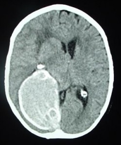
Throughout the week my confidence grew, and the staff helped me relax, it felt easy to fit into the department. By the end of the week, I had performed eight CT head examinations and two abdomen/pelvis examinations. There are many reasons that head CT scans are performed. A few of the more common reasons are to detect brain injuries through trauma such as fractures of the skull or bleeds in the brain, another common referral for head CT examination are to detect bleeding due to a ruptured aneurysm or blood clots in the event of strokes.
I found utilising CT to look for pathologies extremely interesting although, due to its fast pace, there is limited amount of time to examine and study the images carefully. I also felt that, unless you were able to canulate patients and administer IV contrast, CT scanning could become very repetitive. It has very clear advantages from the patients’ point of view, the speed and ease of the examination enables patients to tolerate examination even through pain and discomfort. Even though some patients are on beds and may need to be manually transferred with the use of a PAT slide on to the CT table top, overall CT examinations seem to be well tolerated.
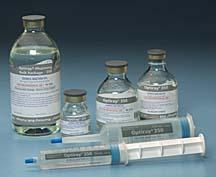
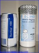
Throughout the week there were many examinations which required IV contrast. Contrast agents (which are usually an iodine compound) used in CT are available in several different forms, some of the more common contrast agents used are, Iodine, Barium, Barium sulfate and Gastrografin. These can be administered in different ways; intravenous injection, oral administration and rectal administration. IV contrast is used in CT is to help highlight blood vessels and to enhance the tissue structure of various organs such as the brain, spine, liver and kidneys.
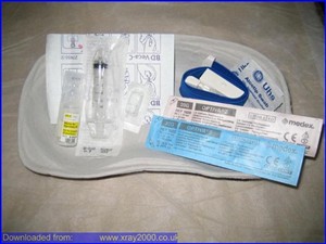
Patients requiring any contrast agent for examinations require safety checks to be carried out. This is to determine any conditions such as diabetes, asthma or any allergic reactions they may have experienced in the past. They are then required to sign an “informed consent form” prior to having their contrast administered and CT exam. This form outlines the potential side effects of the contrast. Some patients experience mild side effects from the contrast agent such as a warm or hot “flush” during the actual injection, a “metallic” taste in the mouth, which usually lasts less than a minute and a sensation like they have to urinate. The patients are told about these potential side-effects and are reassured that these sensations quickly subside. Experiences vary depending on the type of contrast used, the rate at which it is administered and individual patient sensitivity. Milder reactions that may take place following the administration of contrast is itching over various parts of the body with hives, lasting from several minutes to several hours after the injection. This type of reaction is usually treated with medication. A more serious reaction, although much less likely, may include breathing difficulty, swelling of the throat, or swelling of other parts of the body. These reactions can be more serious if not treated immediately.
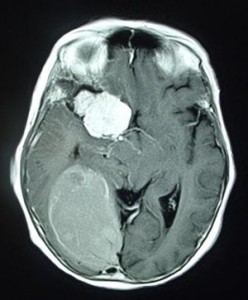
Overall my week in CT was interesting and very fast paced. I am extremely happy to have achieved my competences and enjoyed learning on new equipment. Attached to this piece of reflection is an image of a CT brain taken from,
http://www.radiologyinfo.org.


 Images taken from; http://www.e-radiography.net/index.htm, showing various contrast used in CT imaging.


contrast for CT

Acetabular Fratures pdf

Tuesday, January 4th, 2011

acetabular fractures

Adrenaline used in surgery pdf

Tuesday, January 4th, 2011

Adrenaline used in surgery

?>