Week 6 Out of Hours Year 3
This week’s placement was my out of hour’s week at Queen Margaret Hospital. I had been really looking forward to this week as I knew there were just two other radiographers working and I was hoping I would have a better chance of being involved with more of the trauma patients arriving in the department. This would give me the opportunity to deal with more challenging situations and the experience of adapting my technique.
Apart from the week having some quiet spells I had quite a few situations to deal with which required me to adapt my technique. I was also able to experience how the department works at night. I found this to be very exciting and I really loved my time in the department. I had advised the radiographers that I had been really looking forward to this week and was hoping to gain as much experience as possible in dealing with any trauma situations.
The first situation I was given was the chance to deal with was an elderly patient with a suspected fractured neck of femur (# NOF). There are a high number of elderly patients attending this department with this type of suspected injury due to the hospital being the main trauma centre. This type of injury has an increasing incidence with age thought to be due to a bone density loss and is more common in elderly females. This is usually due to a lack of oestrogen, which is common due to the menopause. Bone density loss can also been seen in patients taking a variety of medications, such as corticosteroids and thyroxine, with injury mostly due to only minor trauma. Classical features for this type of injury are the patients leg is usually shortened and externally rotated and pain on rotation and tenderness over the femoral neck.
The protocol for views needed at Queen Margaret for trauma situations dealing with suspected #NOF are an anterioposterior (AP) pelvis view and an air gap technique for the lateral (Lat) view. I had previously assisted in this situation before while on placement here, but had never been given the opportunity to perform these views. I had mixed emotions, excited, nervous and very cautious. I was comfortable with the radiographer I was working alongside knowing that they would allow me to proceed with the examination under supervision. I also knew they trusted me not to proceed with the exposure without them checking my positioning was correct first.
The AP pelvis was relatively straight forward although it can be difficult sometimes to see if all the correct anatomy will be on the cassette as patients tend not to lie in the middle of the trolley. I took my time and obtained an AP pelvis with all the relevant anatomy included for a #NOF, which was confirmed on viewing. I then positioned for the horizontal beam lateral (HBL), making sure everything was lined up as I thought it should be and then got the radiographer to check my positioning. I took the exposure and processed the image. On observing the image it was apparent that I could have centred down just a few more centimetres; however, the image showed clearly that the angulation of the head of femur was not in the correct position and the patient’s femur was displaced. We went on to repeat the HBL image which gave me the opportunity to repeat the examination and correct my positioning for a perfect image.
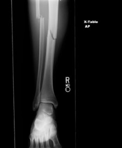
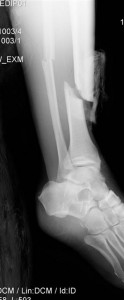
I was also able to visit resus on a few occasions, one occasion being for a gentleman who had fallen down the stairs and had injured has lower right leg. The patient had previous x-rays on his leg as he had a history of Osteomyelitis. This was my first time in resus in a long time so while the doctor was finishing his discussion with the patient the radiographer familiarised me again with the department. I had forgotten that they performed all the examinations with a mobile machine and had been expecting to be using a ceiling mounted tube. Realising this added to my nerves as I immediately thought the examinations were going to be difficult to achieve.
I had observed the patient while I entered the department and had observed that he was covered in blood and his leg was in an unnatural position. I had been advised a fractured tibia is usually associated with a fractured fibula and deformities are common. Deformities or angulation may be obvious on observation due to the foot being abnormally rotated. I obtained the request card to find out what views the doctor was requesting and advised the radiographer what views I thought would be the most appropriate projections.
The doctor had requested an AP and lateral views of the patient’s tibia and fibula from the knee down an AP foot and an AP hand. The most difficult part of the examinations was obtaining the distance for the lateral tib/fib, due to the room size. Moving the patient’s leg for positioning was relatively easy as he had self-anesthetised on alcohol.
I found both examinations very exciting to perform. They were new and challenging situations which I really enjoyed. On the whole the department was quiet due to my shifts being evenings and weekend. I felt I had more of an opportunity to relax and perform more challenging examinations without the pressure of a busy department, although I do feel one week is not enough. I would personally like more of an opportunity to do more out of hours work.
Attached to this piece of writing are images of fractured neck of femur and tib/fib.

 Print This Post
