Archive for December, 2009

Week 8 Elective Placement Year 3

Tuesday, December 22nd, 2009

This week I had been allocated to the rooms that deal specifically with orthopaedic clinics and walk-in GP patients. I have performed many images for the orthopaedic clinics and I feel my ability and skills have grown throughout the week.

There have been audit and review clinics all week and I have had lots of hands on experience. As well as my skills improving I feel I have established a good routine. This is something that I have been working towards for quite a while and it is good to finally have the consistency in my work that a good routine brings. I hope that I will be able to maintain this routine and carry it forward with me on my next placement.

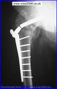
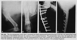
One of the more interesting experiences I have had during this placement involved a patient who had been referred to the department from an orthopaedic clinic. This patient had undergone an internal fixation to her left humerus due to a comminuted fracture 3 weeks previously but the consultant was questioning the alignment of the humerus. A comminuted fracture of a bone is where the fractured bone is in three or more pieces.

The patient was very frightened and was experiencing a large degree of pain. Due to this pain she was unwilling to move her arm. I was able to persuade her by explaining the procedure first and allowing her to manoeuvre her arm by herself; I did however advise her I would help if she needed assistance. She was understandably reluctant to remove her arm from the sling that it was in. I explained I had to take her arm out of the sling to obtain the views needed for her consultant.

To obtain the anterioposterier (AP) view I placed a set of steps with a high support handle attached to them to the left side of the patient, this allowed her to support the weight of her lower arm while she stood in position for the AP view. I explained she would be able to support her arm for the next view by placing her hand on her tummy and holding her arm away from her body. Lifting her arm from the support caused her to cry out in pain. I heard what sounded like a loose screw in the metal and a crepitus sound.

On viewing the images it was clear that 3 of the screws had dislodged from the metal work and away from the bone and the patients arm was not fixed into position. You were also able to view the segments of bone on the images which were in three separate parts. The patient was instructed to return to the orthopaedic clinic for remedial procedures.

Before leaving Perth I managed to follow up on the patient to see if she returned to theatre. The patient had been admitted to one of the wards and was scheduled to return to theatre in the next day.

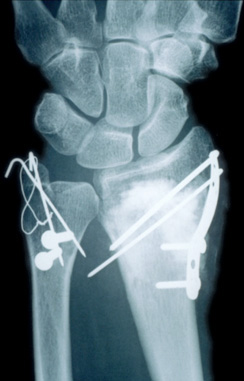
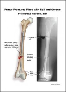
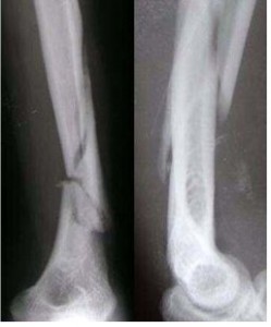
Attached to this piece of writing are images of comminuted humerus fracture and fixations. The first image resembles my patient fractured humerus.


Week 7 Elective Placement Year 3

Tuesday, December 22nd, 2009

This was my first week of a two week elective placement. On arrival to the department I was introduced to staff and shown round by the superintendent. I had been looking forward to this placement and was happy with the schedule that had been arranged for me during my visit. I had specifically requested I would like some hands on with any A & E patients.

It took me a few days to familiarise myself with the systems and equipment, as every room had different types. The department has designated areas for different patients. For my first week I was placed in the rooms which are specifically for A & E and ward patients. I found it all a bit overwhelming at first as the radiographers do almost everything. When a patient arrives from A & E or a ward, the radiographer enters their details and examinations required into the system, after checking their previous history and justifying the examination. They then proceed with the request and then post process the details when finished. Every exposure taken is recorded in detail. They are written on the back of the patients’ request card, the card is then scanned into the system. The exposures are recorded once again during the post processing.
I expected things to be different here but I wasn’t quite prepared for just how different everything was. The equipment here is quite dated such as a Siemens Polydoros 80s which, luckily, I have used previously.

Protocols at the Perth Royal Infirmary are also very different as each orthopaedic surgeon has their own preferences of what views they would like for each examination. So if you have a patient who has had a knee replacement you have to refer to the consultants’ lists and see which views he requests before you perform the examination. Once I understood the procedures and protocols I found this to be a very good system of working.

The A&E patient list wasn’t as exciting as I initially thought. However there were lots of post operative patients arriving from the wards and this gave me the opportunity to practice horizontal beam laterals (HBL). In the past I haven’t had much of an opportunity to have hands on practice for this type of examination. It is performed at Queen Margaret Hospital, using the air gap technique with the up-right bucky for trauma patients with a suspected neck of femur fracture. Here they perform their HBL without the air gap technique but the principals are the same. This examination is performed on patients who are post operative, trauma and some orthopaedic clinic reviews. I was given the chance to perform a few and through practice I found them not as difficult as I initially thought.

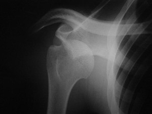
My other experience this week was performing an examination on a patient coming in through A&E with a suspected dislocated shoulder. The examination was quite difficult due to the patients’ pain, although the pain relief began to work and I was able to assist with the views needed. Once the examination was finished I was allowed to return with the patient to the resuscitation unit at A&E and observe while the doctor manipulated the shoulder back into position. It took a doctor and 2 nurses to perform the manipulation. While the doctor was pulling the patients arm down and away from the patients body, one nurse has a bed sheet placed around the top of the patients arm to pull the shoulder up, with the other nurse held the patient on the bed. After a few minutes of pulling, you were able to hear the patients arm slide back into position.

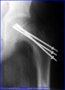
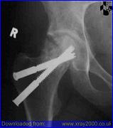
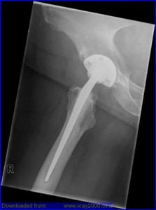
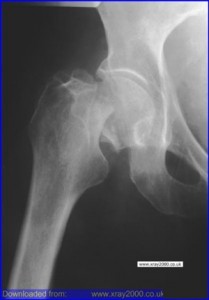
Attached to this piece of writing are images of fractured neck of femur and a hip replacement. A patient with a suspected fracture or a new replacement requires a HBL to show positioning of the fracture or replacement. There are also images of shoulder dislocations.


Week 6 Out of Hours Year 3

Sunday, December 6th, 2009

This week’s placement was my out of hour’s week at Queen Margaret Hospital. I had been really looking forward to this week as I knew there were just two other radiographers working and I was hoping I would have a better chance of being involved with more of the trauma patients arriving in the department. This would give me the opportunity to deal with more challenging situations and the experience of adapting my technique.

Apart from the week having some quiet spells I had quite a few situations to deal with which required me to adapt my technique. I was also able to experience how the department works at night. I found this to be very exciting and I really loved my time in the department. I had advised the radiographers that I had been really looking forward to this week and was hoping to gain as much experience as possible in dealing with any trauma situations.

The first situation I was given was the chance to deal with was an elderly patient with a suspected fractured neck of femur (# NOF). There are a high number of elderly patients attending this department with this type of suspected injury due to the hospital being the main trauma centre. This type of injury has an increasing incidence with age thought to be due to a bone density loss and is more common in elderly females. This is usually due to a lack of oestrogen, which is common due to the menopause. Bone density loss can also been seen in patients taking a variety of medications, such as corticosteroids and thyroxine, with injury mostly due to only minor trauma. Classical features for this type of injury are the patients leg is usually shortened and externally rotated and pain on rotation and tenderness over the femoral neck.

The protocol for views needed at Queen Margaret for trauma situations dealing with suspected #NOF are an anterioposterior (AP) pelvis view and an air gap technique for the lateral (Lat) view. I had previously assisted in this situation before while on placement here, but had never been given the opportunity to perform these views. I had mixed emotions, excited, nervous and very cautious. I was comfortable with the radiographer I was working alongside knowing that they would allow me to proceed with the examination under supervision. I also knew they trusted me not to proceed with the exposure without them checking my positioning was correct first.

The AP pelvis was relatively straight forward although it can be difficult sometimes to see if all the correct anatomy will be on the cassette as patients tend not to lie in the middle of the trolley. I took my time and obtained an AP pelvis with all the relevant anatomy included for a #NOF, which was confirmed on viewing. I then positioned for the horizontal beam lateral (HBL), making sure everything was lined up as I thought it should be and then got the radiographer to check my positioning. I took the exposure and processed the image. On observing the image it was apparent that I could have centred down just a few more centimetres; however, the image showed clearly that the angulation of the head of femur was not in the correct position and the patient’s femur was displaced. We went on to repeat the HBL image which gave me the opportunity to repeat the examination and correct my positioning for a perfect image.

I was also able to visit resus on a few occasions, one occasion being for a gentleman who had fallen down the stairs and had injured has lower right leg. The patient had previous x-rays on his leg as he had a history of Osteomyelitis. This was my first time in resus in a long time so while the doctor was finishing his discussion with the patient the radiographer familiarised me again with the department. I had forgotten that they performed all the examinations with a mobile machine and had been expecting to be using a ceiling mounted tube. Realising this added to my nerves as I immediately thought the examinations were going to be difficult to achieve.

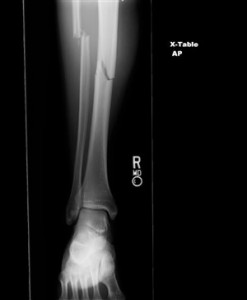
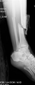
I had observed the patient while I entered the department and had observed that he was covered in blood and his leg was in an unnatural position. I had been advised a fractured tibia is usually associated with a fractured fibula and deformities are common. Deformities or angulation may be obvious on observation due to the foot being abnormally rotated. I obtained the request card to find out what views the doctor was requesting and advised the radiographer what views I thought would be the most appropriate projections.

The doctor had requested an AP and lateral views of the patient’s tibia and fibula from the knee down an AP foot and an AP hand. The most difficult part of the examinations was obtaining the distance for the lateral tib/fib, due to the room size. Moving the patient’s leg for positioning was relatively easy as he had self-anesthetised on alcohol.

I found both examinations very exciting to perform. They were new and challenging situations which I really enjoyed. On the whole the department was quiet due to my shifts being evenings and weekend. I felt I had more of an opportunity to relax and perform more challenging examinations without the pressure of a busy department, although I do feel one week is not enough. I would personally like more of an opportunity to do more out of hours work.

Attached to this piece of writing are images of fractured neck of femur and tib/fib.


?>