Archive for February, 2009

Week 9 Year2

Friday, February 27th, 2009

On clinical placement this week, as part of my continual learning, I went on a number of mobile x-ray examinations. I had helped perform a few previously and although mobile x-rays have their own inherent challenges, I enjoyed the experience. However this week was exceptionally difficult for a number of reasons.

One of the biggest challenges was seeing some of the conditions the patients were suffering from. Doctors only generally request a mobile examination if the patient is extremely unwell and unable to travel to the department. This is due
to radiation protection issues but also because the image from a mobile x-ray is not of the same quality as a static machine. However I don’t think I was prepared for just how unwell some of these patients were.

Among the wards I attended were the oncology ward, theatre recovery and the high dependency unit. I don’t think anything can prepare you for seeing some of the patients’ conditions and illnesses and therefore the difficulty is maintaining your professionalism. Trying to not look shocked or frightened and keeping composed whilst performing the x-ray. I tend to talk a lot when I’m nervous and found this to be an advantage as it helped to both relax the patient and myself.

Another challenge I had to overcome was positioning the cassette under the patient when they had numerous tubes and wires attached to them. Also removing the cassette after the exposure and trying to take care not to cause any pain
or discomfort to the patient. This was made easier in cases where I went to theatre recovery or the high dependence unit as there were nurses there to help manoeuvre the patient in order to facilitate positioning the cassette.

Mobile x-rays are not always straight forward, and the radiographer needs to be capable of problem solving. For example, some considerations are, is the patient in a small room or on a ward? In the case of a patient being in a small room you may have to position the cassette before bringing in the machine. Once the machine is in the room, it can be difficult positioning the tube correctly, and at a required distance for you to achieve the best possible image. One difficulty I ncountered in achieving distance was solved by lowering the patient’s bed to achieve a greater distance for the image.

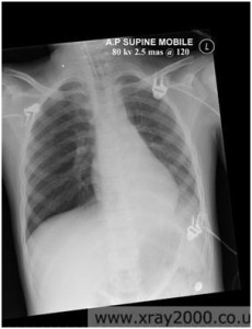
I enjoyed my experience and feel I gained a lot of practical knowledge in patient management. Attached to this piece of writing is an image of a good portable chest x-ray, it highlights the fact that the quality can be diagnostic, however the image is not as good as one performed on a static machine.


Week 8 Year 2

Thursday, February 19th, 2009

Journal

This week on placement I have been at the Royal Victoria Hospital. The hospital is part of the Western General and is dedicated to care of the elderly and an open access service for GP patients and clinics.

I encountered a few challenges throughout the week, these ranged from transferring uncooperative patients and adapting technique to allow for patients conditions. Some patients encountered fear due to suffering pain on movement or weight bearing on fractures. I also encountered many patients with limited or no range of movement. In these instances I had to adapt my technique to obtain the best possible image.

Some patients were a challenge when helping them transfer from their chairs to the table. I found a lot of patients wanted the staff to lift them rather than them transferring themselves on their own.

There was a particular case which I found to be challenging; this involved a patient attending the department from a ward for an ankle x-ray. The patient was brought down to the department in a chair. After confirming all the details I moved the chair to the side of the table. The patient informed me she was frightened to stand, so I advised her to put most of her weight on her good leg while transferring. Getting assistance I assured her we would help her on to her feet and be by her side while transferring her from her chair. On helping her to her feet she seemed to use little effort to support herself making us support most of the weight. After a difficult transfer I then had to try and achieve a good projection. The patient suffered with oedema in her legs and ankles and found it difficult to move them, making positioning difficult.

The AP projection wasn’t too difficult to obtain using the little toe as a marker. However the lateral projection caused a few problems. The patient couldn’t turn onto her side so I then had try and turn her with the use of a pad to hold her in position. It was difficult to see if the patient’s leg was in the correct position due to the swelling but I was able to achieve a good projection.

Week 7 Year 2

Monday, February 16th, 2009

Journal

This week on placement I had a number of patients from an oncology clinic for follow up chest x-rays. These patients were all referred due to having conditions called seminoma and teratoma. Both these conditions are cellular cancers which
started, in these cases, in the testes. Seminoma is a radiosensitive malignant neoplasm of the testis, and teratoma is a germ cell tumour composed of multiple cell types derived from one or more of the 3 germ layers.

According to emedicine, 3% of testicular teratomas are known to metastasize in adults and adolescents. There are two types of teratomas, mature and immature. Mature teratomas are usually found in women and are usually found to be benign, whereas the immature teratomas are usually found to be malignant and more commonly found in males. Teratomas are thought to be congenital, but are often not diagnosed until later in life.

The patients had all been referred for follow up chest x-rays by their consultant to check for any metastases in the lungs. I had never heard of any of these diseases before and looking over the request cards I noticed all the patients were all young men, between the ages of twenty five and thirty five. After some research into these diseases I discovered they were both cellular cancers, beginning in the embryonic stage. Once discovered, these diseases are monitored carefully as there is the possibility for them to metastasize.

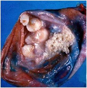
All patients x-rayed had no obvious signs of metastases and were due to see their consultant after their x-ray. Attached to this piece of writing are images and a website I used to research these diseases. Throughout my research I found out both male and females can have this condition, however in this case, all patients were male.

Both seminoma and teratmoas can be found in different parts of the body. According to emedicine, the most common location is sacrococcygeal. As they arise from totipotential cells, they are encountered commonly in the gonads.
The most common location being the ovary, and occurring less frequently in the testes. Occasionally teratomas occur in midline embryonic cell rests and can be mediastial, retroperitoneal, cervical, and intracranial. Cells differentiate along various germ lines, essentially recapitulating any tissue of the body. Examples include hair, teeth, fat, skin, muscle, and endocrine tissue.

Testicular cancer treatment involves radical surgical. This surgery involves the removal of the testicle. As part of the diagnosis of testicular teratoma, tissue diagnosis and distinction from other forms of testicular cancer is important in subsequent management. Regional lymph nodes may also be sampled or removed during surgery. Teratoma of the testicle is relatively resistant to radiotherapy but responds well to chemotherapy as an additional testicular cancer treatment to surgery or as a primary treatment in advanced disease.

Following the surgery or chemotherapy the patient will be closely followed to detect any recurrences early. Follow up includes self-testicular examination, measurement of tumour markers in the blood, serial chest x-ray and regular
abdominal and thoracic CT scanning.


Week 6 Year 2

Friday, February 13th, 2009

Journal

This week I have been on placement in the Western General hospital. This was my first time in this particular hospital so I was naturally a bit apprehensive, not knowing where to go, or knowing any of the staff. However these fears were soon
allayed and, in the end, I really enjoyed it. The staff were lovely, so friendly and helpful and it wasn’t long before I relaxed into my placement. This allowed me to concentrate on familiarising myself with the equipment and finding out how the department runs.

I was curious to see that they performed chest x-rays with the upright bucky. Previously I had only seen a chest x-ray performed with the cassette placed outside the bucky without the use of a grid. I wondered why they chose to perform the examination this way and also how they could be sure they were going to get the entire chest on the image.

After observing this technique a few times, I began to understand the difference between placing the cassette inside or outside of the bucky. The radiographer showed me how the cassette is centred to the bucky, so this gave the radiographer a reference with which to firstly position the patient and then collimate the beam. Once I performed a chest x-ray using this technique I found it quite straight forward.

After performing the examination, I noticed the exposure set on the console seemed very high. When using the bucky for the examination, there is then a need to increase the KVp, which I thought would increase patient dose. I couldn’t understand how this technique could conform to the ‘low as reasonably practicable’ rule regarding radiation, as set out by IRMER 2000 (Ionising Radiation (Medical Exposure) Regulations). However it was explained that this was the radiologists request as this would allow him to see behind the mediastinum and heart for any pathology.

I have since looked at two chest x-rays, one of which I performed using the high KVp and in the bucky, and one taken out of the bucky using the lower KVp from a previous hospital. On comparison, in my opinion, it was possible to see much more detail in the image using the higher KVp and the grid. There is better detail in the lungs and behind the heart as well as through the spine. I found this an interesting method and at some point would like to investigate this method further. I have done some research and since found out the technique used at the hospital can be used to reduce exposure error. An
iontomat is used in collaboration with a high KVp and a grid, while using the smallest possible mAs, therefore reducing patient dose. I have included a link for a website I found while researching the different exposures and techniques,
which gives examples of images at different exposures.

?>